Examine skin and select an area that appears abnormal. Cut a small piece of the skin and place it into a drop of saline on a clean glass slide. Cover with a slip cover, and additional saline if needed.
Compare your sample with the following Disease Vectors:
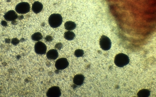
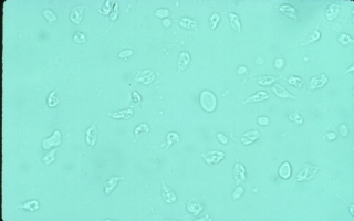
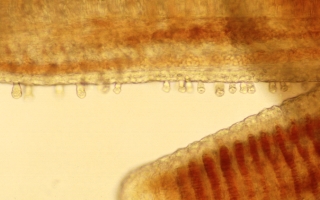
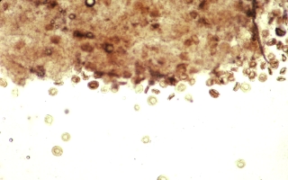
Amyloodinium sp 0.03 to 0.05 mm spherical
to pear shaped and attached by a short stalk. blackish in color and vacuolated
(e.g. full of tiny spherical bubbles) under transmitted light
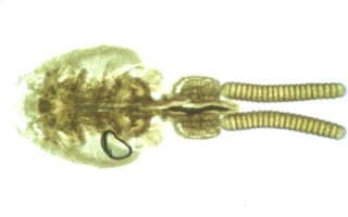
Caligus sp. a parasitic copepod 2 to 3 mm in length
(visable to the naked eye)
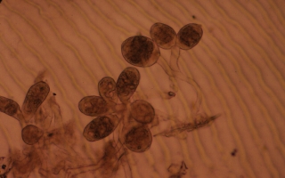
Epistylis sp., a stalked colonial protozoan.
0.05 mm cup or tulip shaped. attached by branching stalks to the skin. cilia
around the rim of the open (apical) end of the "bell. May contract to a
ball-shape or detach from stalks and swim away. colonies are large enough
to be visible with the naked eye as soft, fluffy growths protruding from
sores on the skin.
Ichthyobodo sp. (Costia), a flagellated
ectoparasite. VERY small, about 0.007 mm or the size of a red blood cell.
It may be found attached to gill or skin cells by its narrow end or moving
in a jerky circular motion in the wet-mount fluid near the tissue
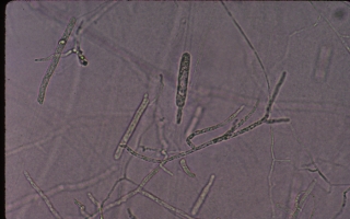
Saprolegnia spp. fungi at 100X to 400X appears
as a branching tangle of root-like filaments (e.g. the hyphae). The filaments
are clear and about 0.005 mm in diameter.
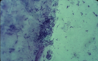
Myxobacteria long (0.003-0.01 mm) thin (0.0005
mm) rod shaped bacteria often seen in clumps or "stacks" which move by flexing
or gliding.
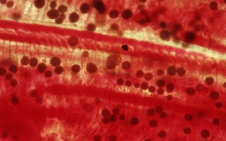
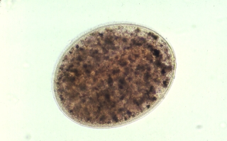
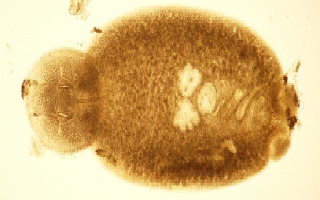
Ichthyophthirius multifiliis ("Ich", marine: Cryptocaryon
irritans), a protozoan ectoparasite. can vary in size from 0.02 mm to nearly
1 mm depending on the age of the parasite The cilia along the perimeter of
the organism beats and slowly propels the protozoan forward. Under transmitted
light in wet-mount preparations, Ich appears brownish in color.
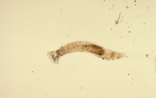
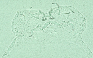
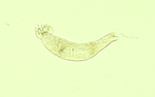
Monogenetic Trematodes parasitic
flatworms. May be small (0.3 to 0.4 mm) or large (2to 3 mm) . Hooks on one
end may be attached to the host fish. If still alive, will flex rapidly or
contract slowly.
Scyphidia sp. a free-living protozoan. stumpy
(~0.04 mm in height), cylindrical shaped with a ring of cilia around the
apical end and an expanded sucker shaped basal end in contact with the skin
of the fish. when detached from the fish change to a fast moving saucer
shape.
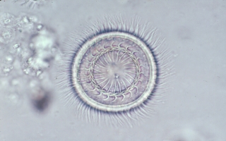
Tricodina sp., a protozoan ectoparasites ~0.04
mm circular saucer shaped and very fast moving when alive. rimmed by cilia
and on the bottom side. peculiar denticular rings with teeth that point
inward.
| file: /Techref/other/pond/tilapia/skin.htm, 3KB, , updated: 2009/7/13 18:44, local time: 2025/9/5 12:13,
216.73.216.166,10-1-199-35:LOG IN
|
| ©2025 These pages are served without commercial sponsorship. (No popup ads, etc...).Bandwidth abuse increases hosting cost forcing sponsorship or shutdown. This server aggressively defends against automated copying for any reason including offline viewing, duplication, etc... Please respect this requirement and DO NOT RIP THIS SITE. Questions? <A HREF="http://www.massmind.org/techref/other/pond/tilapia/skin.htm"> Tilapia Topic: Skin Microscopy</A> |
| Did you find what you needed? |
Welcome to massmind.org! |
Welcome to www.massmind.org! |
.